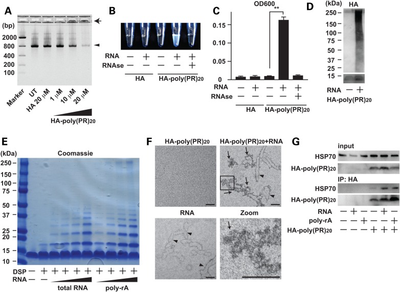
Figure 4.
Poly-PR peptide interacts with RNA and forms aggregates. (A) Agarose gel electrophoresis of solutions containing GFP mRNA with HA peptide or HA-poly-(PR)20 peptide at indicated concentrations. An arrowhead shows GFP mRNA and an arrow shows insoluble RNA aggregate. (B and C) The measurement of turbidity using the optical density at 600 nm (OD600) of the solution containing HA peptide or HA-poly-(PR)20 peptides mixed with or without yeast total RNA. The image represents visible formation of poly-PR aggregates that can be reversed by treatment with RNase. N = 3 biological replicates. (D) Immunoblot analysis of HA-poly-(PR)20 peptides mixed with or without yeast total RNA. SDS–PAGE was performed with a 4–20% gradient gel. (E) Coomassie blue staining of poly-PR aggregates induced by yeast total RNA or monopolymeric poly-adenilyc acids (poly-rA). Samples were crosslinked with dithiobis (succinimidyl propionate) (DSP, 2 mm) before applied to SDS–PAGE. (F) Electron microscope images of poly-PR aggregates induced by RNA. The scale bar shows 50 nm. Arrowheads show RNA and arrows show poly-PR aggregate. (G) Co-immunoprecipitation of human recombinant HSP70 with HA-poly-(PR)20 peptides mixed with or without yeast total RNA or poly-rA. Each experiment was repeated at least three times and representative images were shown. Asterisks indicate a significant difference analyzed by one-way ANOVA followed by Dunnett's test (**P < 0.01 and *P < 0.05).

