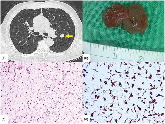
Figure 1.
A, CT revealed a well-circumscribed homogeneous mass in the left upper lobe (yellow arrow). B, The resected tumour was gelatinous and 1.7 × 1.3 × 1 cm. C, Histological examination revealed a hypocellular neoplasm composed of elongated and stellate cells in a myxoid stroma with no mitotic activity, no lipoblasts and delicate blood vessels (haematoxylin and eosin ×200). D, The tumour cells were immunohistochemically positive for vimentin (×400).

