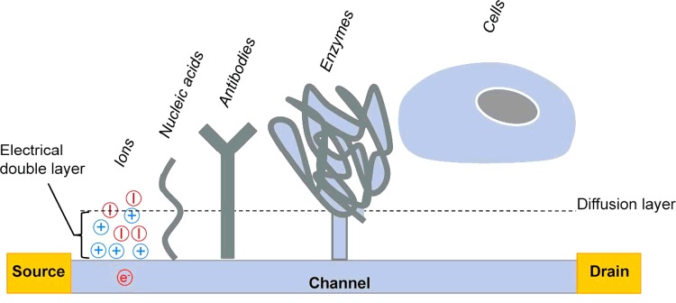
Figure 3. Schematic representation of a biologically sensitive ISFET and different types of analytes with variable sizes in relation to the thickness of the EDL (analyte size is not to scale).
Only DNA molecules present charges inside the EDL, whereas only fractions of the much larger antibody and enzyme molecules directly affect the EDL (only in the case of diluted buffer concentrations). In the case of cells, the cellular membrane is clearly outside this range, since typical average distances between the cellular membrane and the surface are in the range 50–100 nm.

