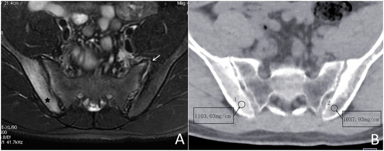
Figure 2.
Patient with axial spondyloarthritis (male, 20 years old, HLA-B27 positive, low back pain for 3 months). (a) Axial T2 weighted shows the abnormal high signal intensity involving subcortical bone marrow of the right ilium (black star) and left sacroiliac joint synovitis (arrow). (b) The water-based image of spectral CT image demonstrates the water concentration of the right ilium (1103.03 mg cm−3) to be higher than that of the left (1037.93 mg cm−3). The synovitis remains CT occult.

