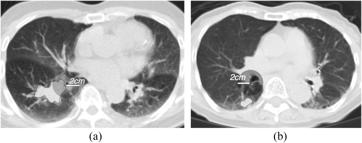
Figure 1.
Representative cases of the central type (a) and peripheral type (b) on CT images. The primary tumour is delineated with a thin black line. The transparent grey circle demonstrates a region 2 cm from the origin of the nearest segmental bronchus. If the margin of the primary tumour is within the circle, it is classified as a central type.

