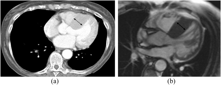
Figure 2.
A 77-year-old female with shortness of breath and palpitations. (a) Standard axial chest CT demonstrated normal lungs and asymmetric hypertrophy of the interventricular septum (arrow, 32 mm). (b) Corresponding cardiac MRI (CMR) confirms asymmetric hypertrophy of the interventricular septum (arrow, 25 mm) consistent with a hypertrophic cardiomyopathy.

