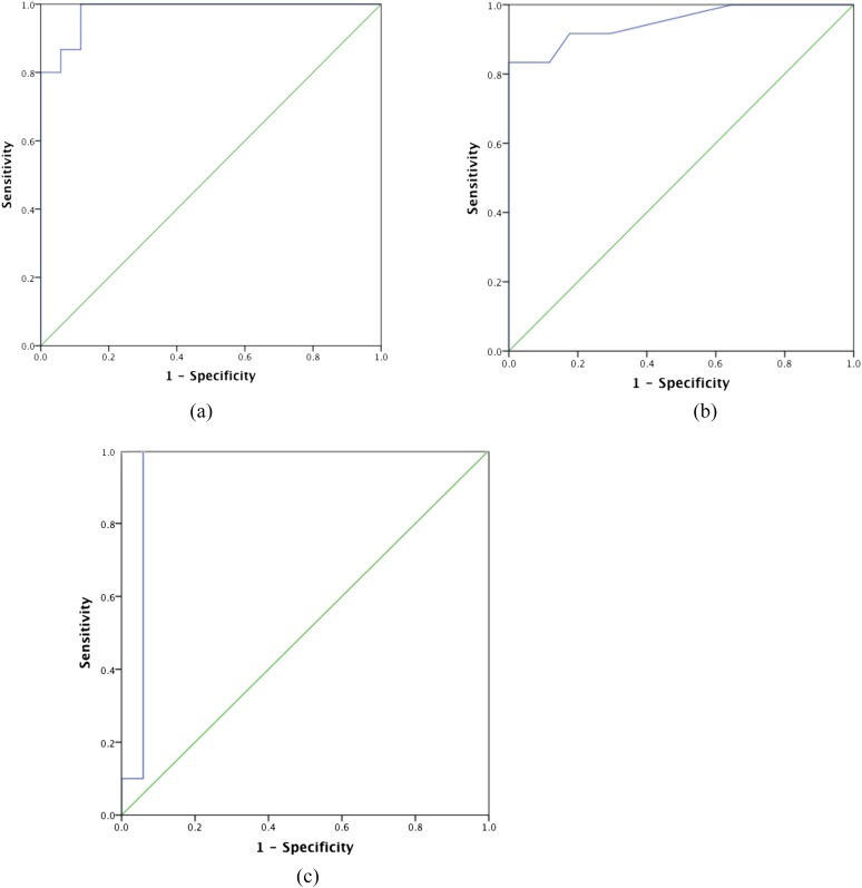
Figure 8.
Receiver-operator curve analysis for (a) left ventricular diameter in dilated cardiomyopathy (DCM), (b) interventricular septal thickness in hypertrophic cardiomyopathy (HCM) and (c) papillary muscle thickness in HCM on standard axial chest CT compared with controls. (a) The area under the curve (AUC) = 0.92, p < 0.0001, using a cut-off value of 47 mm, yielded a sensitivity and specificity of 93% and 88%, respectively. (b) The AUC = 0.95, p < 0.0001, using a cut-off value of 14 mm, yielded a sensitivity and specificity = 83% and 100%, respectively. (c) The AUC = 0.95, p < 0.0001, using a cut-off value of 9 mm, yielded a sensitivity and specificity = 100% and 94%, respectively.

