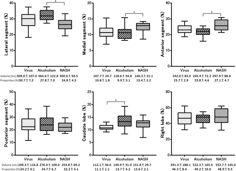
Figure 4.
Box plot showing the proportion of each segment to the total liver volume of the three aetiologies in Child–Pugh Class B. The proportion of lateral segment to the total liver was significantly smaller in patients with non-alcoholic steatohepatitis (NASH) than in those with alcoholic cirrhosis (p < 0.001), the proportion of the medial segment was significantly larger in patients with NASH than in those with alcoholic cirrhosis (p = 0.045), the proportion of the anterior segment was significantly larger in the patients with NASH than in those with alcoholic cirrhosis (p = 0.003) and the proportion of the caudate lobe was significantly smaller in the patients with virus-related cirrhosis than in those with alcoholic liver cirrhosis (p = 0.001). There were no significant differences in the proportion of the posterior segment and right lobe to the total liver among the patients with the three different aetiologies (p > 0.05).

