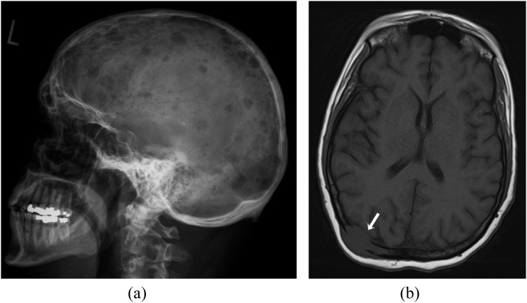
Figure 1.
A 60-year-old female with multiple myeloma. (a) Plain radiograph of the skull (lateral view) demonstrating multiple lytic lesions. (b) Axial T1 weighted MR image showing an expansile lesion involving right parietal bone with extraosseous soft-tissue component (arrow). The lesion shows hypointense signal compared with that of fatty marrow of the diploic cavity.

