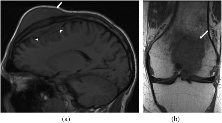
Figure 10.
A 22-year-old male with Burkitt's lymphoma. (a). Sagittal T1 weighted (T1W) MR image showing T1 hypointensity involving the frontal bone with associated extradural (arrowheads) and scalp soft-tissue mass (arrow). (b) Coronal T1W MR image shows hypointense lesion involving the distal end of the femur (arrow).

