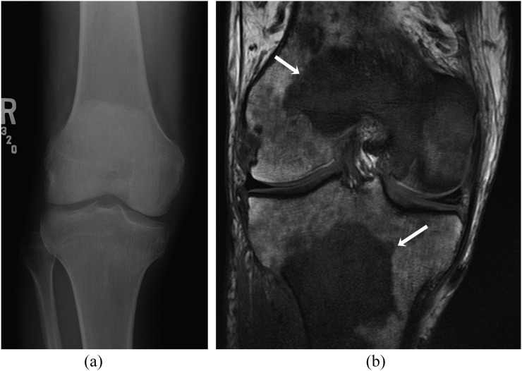
Figure 11.
A 45-year-old male with chronic myeloid leukaemia and multiple myeloid sarcoma. (a) Anteroposterior radiograph of the right femur shows no significant abnormality. (b) Coronal T1 weighted MR image shows large T1 hypointense lesions in the distal femur and proximal tibia (arrows) and numerous other ill-defined smaller lesions.

