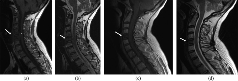
Figure 3.
A 43-year-old female with multiple myeloma. (a, b) Pre-treament sagittal T1 weighted (T1W) (a) and T2 weighted (T2W) (b) MR images of the cervical spine showing marrow involvement of the C5 vertebra with pre-vertebral (arrows) and anterior epidural (arrowhead) soft-tissue components (b). Post-treatment sagittal post-contrast T1W (c) and T2W (d) MR images showing hypointense marrow signal of the affected C5 vertebra (arrows) with resolution of soft-tissue component.

