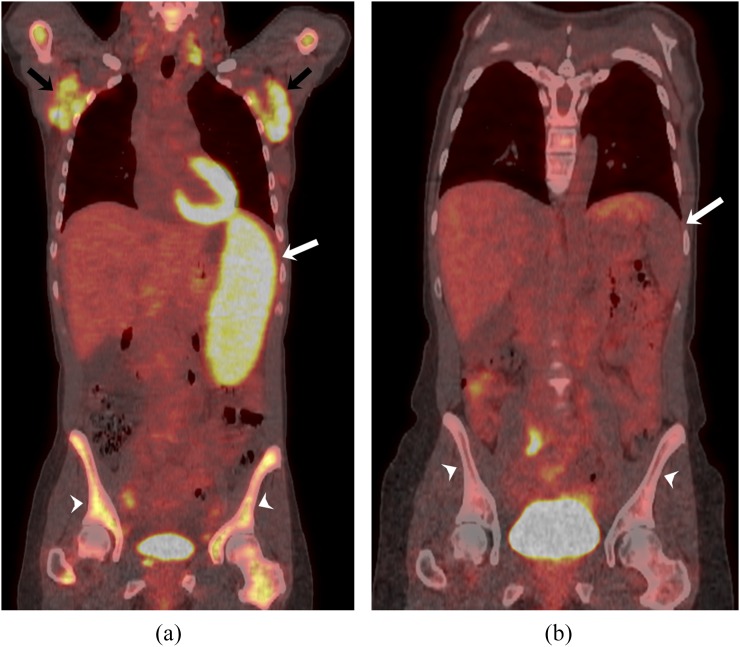
Figure 6.
A 43-year-old female with Waldenström macroglobulinaemia. (a) Coronal-fused fluorine-18 fludeoxyglucose positron emission tomography (18F-FDG PET)/CT image shows 18F-FDG-avid bilateral axillary lymphadenopathy (black arrows), splenic involvement (white arrow) and diffuse skeletal uptake (white arrowheads) consistent with bone marrow involvement. (b) Post-treatment coronal fused 18F-FDG PET/CT image shows significant interval decrease in 18F-FDG-avid disease involving spleen (white arrow) and bone marrow (white arrowheads).

