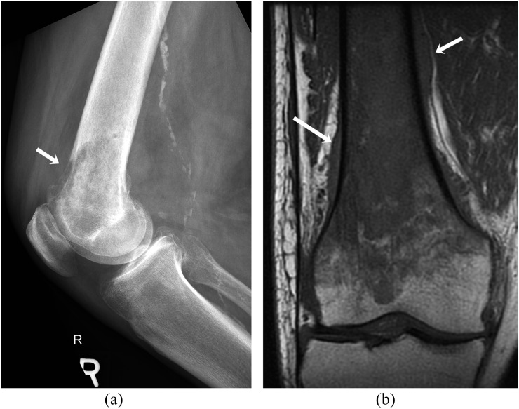
Figure 7.
A 80-year-old female with primary B-cell non-Hodgkin's lymphoma involving the right distal femur. (a) Lateral radiograph of the right femur shows ill-defined lytic lesion involving distal femoral metaphysis with cortical destruction (arrow). (b) Coronal T1 weighted MR image shows low signal intensity of the tumour relative to fatty marrow of the epiphysis and periosteal reaction and soft tissue (arrows).

