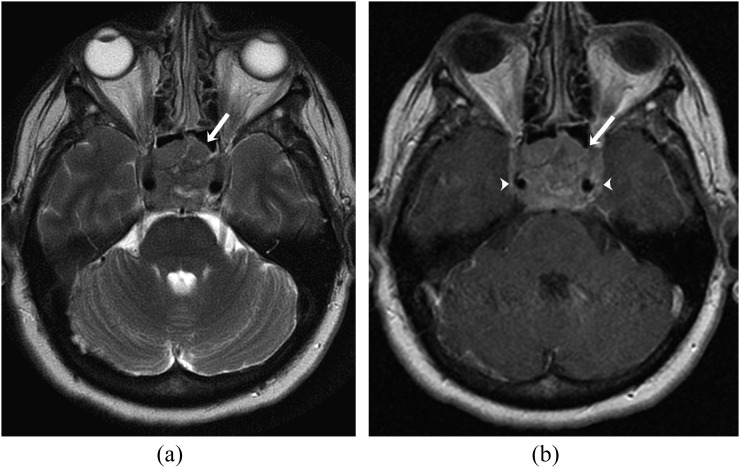
Figure 9.
A 65-year-old female with diffuse large B-cell lymphoma. (a) Axial T2 weighted (b) MR image showing heterogeneous expansile lesion involving the clivus (arrow). (b) Axial contrast-enhanced T1 weighted MR image showing heterogeneous enhancement of the lesion (arrow) with encasement of cavernous segment of internal carotid arteries on both sides (arrowheads).

