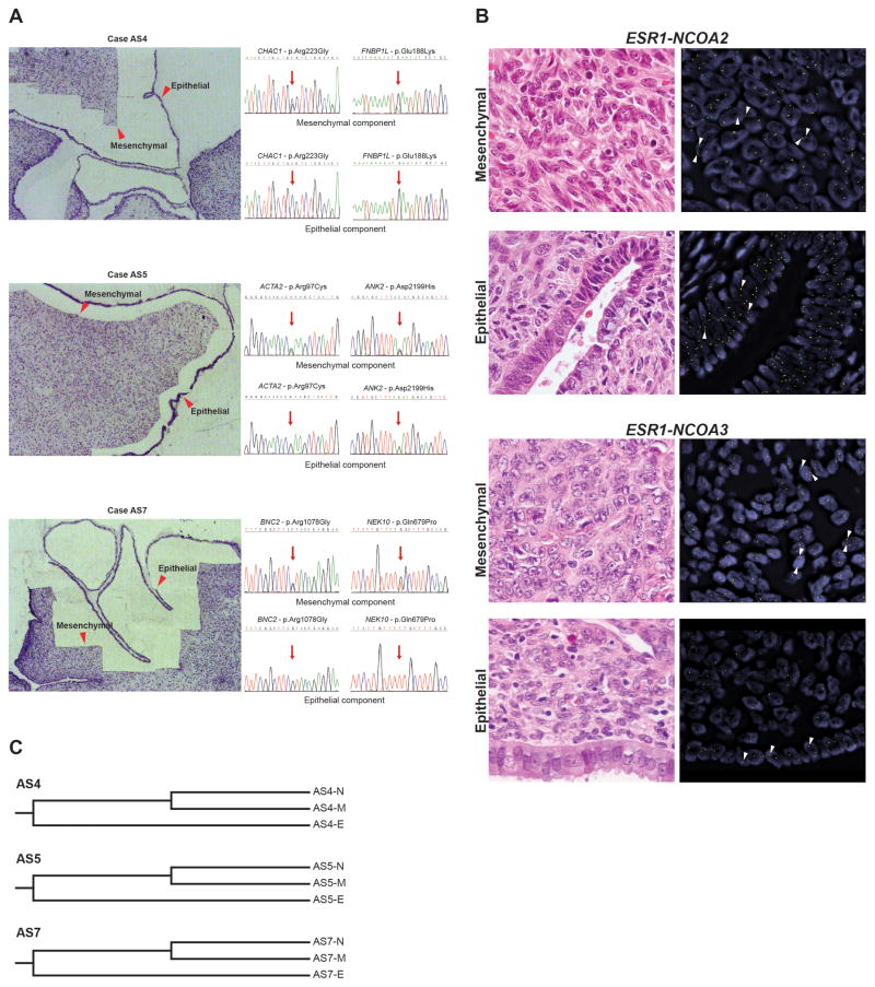
Figure 3. The presence of somatic genetic alterations in the mesenchymal component but not in the epithelial component of uterine adenosarcomas.
A, Representative micrographs of negative microdissection, and the associated Sanger sequencing traces for selected mutations in the separately microdissected mesenchymal and epithelial components of three uterine adenosarcomas. The regions from which DNA was extracted are indicated by red arrows. Note that the mutations were restricted to the mesenchymal components in each case. B, Representative micrographs (haematoxylin and eosin left, fluorescence in situ hybridization (FISH), right) of the mesenchymal and epithelial components of uterine adenosarcomas AS8 (top) and AS4 (bottom). The presence or absence of the rearrangements using break-apart probes are indicated by white arrows. FISH analysis using two-colour break-apart probes for NCOA2 and NCOA3, with 5′ NCOA2 and NCOA3 green, 3′ NCOA2 and NCOA3 orange. Note that NCOA rearrangements are restricted to the mesenchymal components in both cases. C, Phylogenetic clustering of mitochondrial DNA D-loop regions in mesenchymal and epithelial components, and matched normal tissue of 3 cases (AS4, AS5 and AS7). The relative phylogenetic distances between matched normal tissue (N), mesenchymal component (M) and epithelial component (E) were determined using the neighbour-joining method.

