Figure 6.
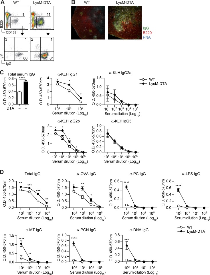
Plasma cells in LysM-DTA iLNs are IgG+ and have higher titers of antigen-specific serum antibodies after immunization. (A) Intracellular staining of iLN plasma cells (B220−CD138+) against IgG and IgM in WT and LysM-DTA iLNs at day14 p.i. (B) Immunofluorescent staining of iLNs to visualize B cell follicles (B220+) and germinal centers (PNA+IgG+). Bars, 100 µm. (C) LysM-DTA and WT mice were immunized with 100 µg KLH in CFA, and serum antibodies were analyzed at day 14. Total serum IgG was diluted 100,000-fold. (D) LysM-DTA and WT mice were immunized with 100 µg OVA in CFA, and serum antibodies were analyzed at day 14. Data in A are representative of three independent experiments. n = 4–5/group. The images in B are representative of four separate mice. Data in C and D are representative of two independent experiments. n = 5/group. Results are mean ± SEM. *, P < 0.05; **, P < 0.01; ***, P < 0.001; ****, P < 0.0001 (unpaired Student’s t test).

