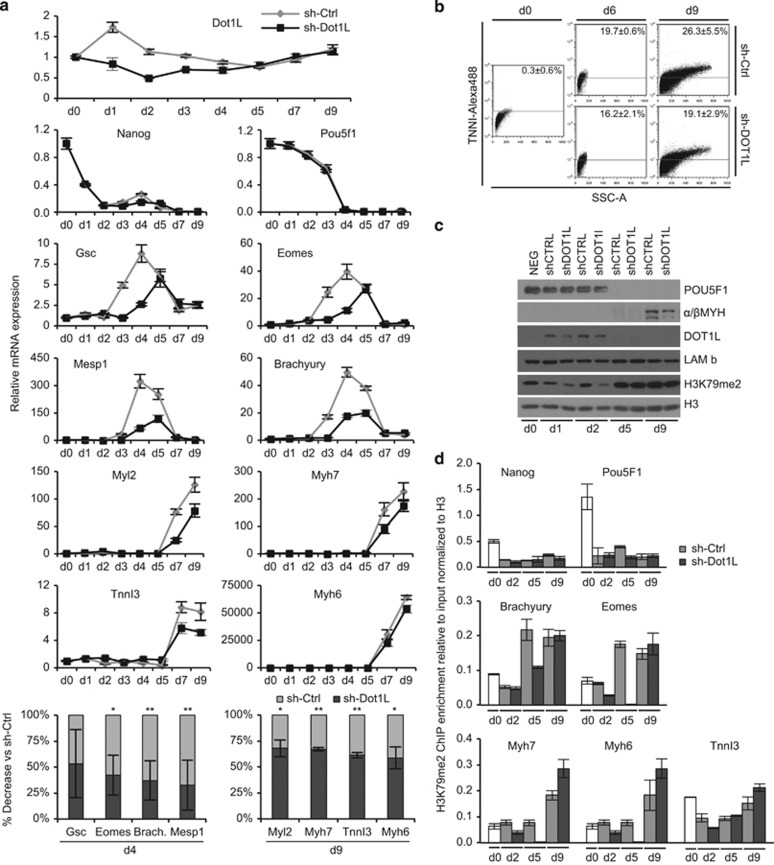
Figure 4.
Disruptor of telomeric silencing 1-like (DOT1L)-mediated di-methylation (me) of H3K79 in cardiogenic differentiation of mouse embryonic stem cells (mES). (a) mRNA levels of Dot1L, pluripotency, mesodermal, and cardiomyocyte (CM)-specific genes in undifferentiated (day 0) mES and after days 1, 2, 3, 4, 5, 7, and 9 of differentiation in control (sh-Ctrl, gray line) and Dot1L-knockdown (sh-Dot1L, black line) cells. Levels are normalized to 18 s or Gapdh, expressed as the mean±S.D. of experimental replicates, and plotted as relative mRNA expression (top). Percentage decrease of Gsc, Eomes, Mesp1, and Brachyury expression at day 4 and Myl2, Myh7, TnnI3, and Myh6 at day 9 in sh-Dot1L versus sh-Ctrl transduced cells. Mean±S.D. of three independent experiments is plotted (**P<0.01; *P<0.05 sh-Dot1L versus sh-Ctrl; bottom). (b) FACS analysis of the percentage of cells expressing TNNI at days 0, 6, and 9 of differentiation in control (sh-Ctrl) and Dot1L-knowkdown (sh-DOT1L) cells. (c) Protein levels, assessed by Western blotting, at days 0, 1, 2, 5, and 9 of differentiation in control (sh-Ctrl) and Dot1L-knockdown (sh-Dot1L) mES. The expression levels for POU5F1, α/βMYH, and DOT1L are normalized to that of lamin B (LAM b), whereas the expression levels of H3K79me2 are normalized to that of unmodified H3. (d) Chromatin immunoprecipitation (ChIP) assay of H3K79me2 binding to the gene locus of Nanog, Pou5F1, Brachyury, Eomes, Myh7, Myh6, and TnnI3 in undifferentiated (day 0) mES (white bars) and at days 2, 5, and 9 after differentiation in control (sh-Ctrl, gray bars) and Dot1L-knockdown (sh-Dot1L, black bars) cells. Levels were determined by qPCR and are expressed as fold change to the input and relative to H3

