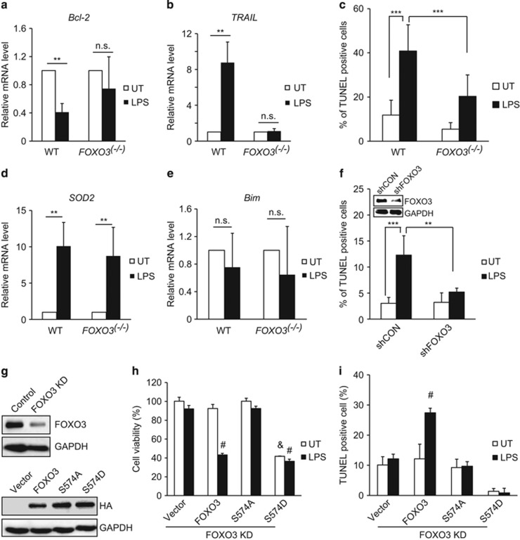
Figure 7.
p-574-FOXO3-dependent macrophage apoptosis. (a and b) Real-time RT-PCR analysis of Bcl-2 and TRAIL mRNA levels in peritoneal macrophages isolated from wild-type (WT) or FOXO3−/− mice and subsequently treated for 24 h with 100 ng/ml LPS (WT n=3, FOXO3−/−n=4). (c) TUNEL assays were performed in peritoneal macrophages isolated from WT and FOXO3−/− mice either untreated (con) or treated with 100 ng/ml LPS for 24 h (n=3 each). (d and e) Real-time RT-PCR analysis of SOD2 and Bim mRNA levels in peritoneal macrophages isolated from WT or FOXO3−/− mice after 24 h of treatment with 100 ng/ml LPS (WT n=3, FOXO3−/−n=4). (f) THP-1 cells were infected with vectors coding for FOXO3-specific shRNA or empty lentiviral particles. After 48 h, cells were either left untreated (UT) or treated with 100 ng/ml LPS for 24 h. TUNEL assays were then performed. Values are mean±S.D. **P<0.01, ***P<0.001, Student's t-test. Inset shows western blot demonstrating magnitude of the knockdown achieved. (g, upper panel) Western blot to assess shRNA knockdown efficiency of FOXO3 in THP-1 cells after 5 μg/ml puromycin selection. (g, lower panel) Western blot in these same cells 48 h after electroporation of HA-tagged WT or mutant FOXO3 proteins. (h and i) Cell viability and TUNEL assay for FOXO3 knockdown cells electroporated with empty vector, HA-WT-FOXO3, HA-FOXO3 S-574A or HA-FOXO3 S-574D. Results are shown after 48 h of either no treatment (UT) or 24 h without treatment followed by an additional 24 h of treatment with 100 ng/ml LPS. Values are mean±S.D. #P<0.001 versus vector/UT, &P<0.001 versus vector/LPS

