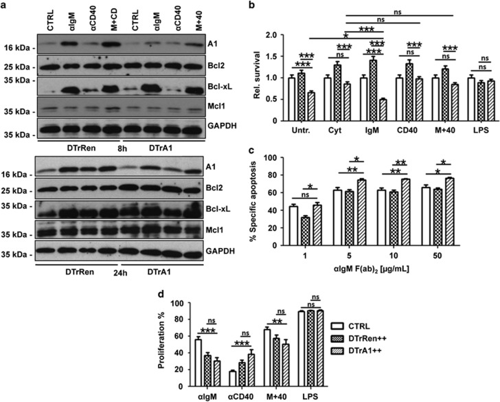
Figure 6.
A1 knockdown sensitizes mature B cells to apoptosis. (a) Quantitation of A1 knockdown efficacy at the protein level in sorted B220+ GFP++ splenocytes derived from mice of the indicated genotypes kept on doxycycline for 17 days. Cells were cultured in the presence of IL-2, -4, -5 alone (CTRL) or stimulated additionally with 1 μg/ml anti-mouse IgM F(ab')2 fragments (αIgM), 1 μg/ml anti-mouse CD40 mAb (αCD40) or both (M+40) in the presence of 1 μg/ml doxycycline for 8 or 24 h. Membranes were probed with antibodies specific for the indicated Bcl2 family members to assess possible compensatory effects or anti-GAPDH to control for protein loading (one out of two independent experiments yielding similar results is shown). (b) Total spleen cells from mice kept on doxycycline were isolated and incubated in media alone (untreated), cytokines (CYT) or cytokines with the indicated mitogens. Cultures were maintained in the presence of doxycycline (1 μg/ml), which was re-added after 40 h. After 72 h cells were stained with antibodies recognizing IgM or IgD and the percentages of viable (Annexin V-) GFP++ IgM+IgD+ B cells was assessed and compared with the percentage of B cells detected straight after killing. As these percentages differ (see also Figure 5), the bar graphs show the ratio of the percentage of IgM+IgD+ B cells in the culture on day 3, divided by the percentage of the IgM+IgD+ B cells present on day 0. Bars represent means±S.E.M. (wt and single transgenic controls n=11, DTrRen n=7, DTrA1 n=9). (c) GFP++ mature splenic B cells (IgMloIgDhi) from DTrA1 or DTrRen mice were sorted and cultured in the presence of graded concentrations of plate-bound anti-IgM F(ab)2 fragments in the presence of 1 μg/ml doxycycline. Cell viability was assessed after 20 h by Annexin V staining. The extent of apoptosis induced specifically by BCR ligation or SYK inhibition was calculated by the following equation: (induced apoptosis/spontaneous cell death) × 100. All experiments were performed in duplicates. Bars represent means±S.E.M. (wt and single transgenic controls n=5, DTrRen n=3, DTrA1 n≥5). (d) GFP++ B cells were cultured in media containing doxycycline (replenished after 40 h) plus cytokines, or cytokines plus the indicated mitogens. Loss of CPD labeling was assessed after 72 h to calculate the percentage of B-cell proliferation (wt and single transgenic controls n=12, DTrRen n=10, DTrA1 n=9). ANOVA followed by Bonferroni post-hoc test was performed to evaluate results for significant differences. *P≤0.05, **P≤0.01, ***P ≤0.001

