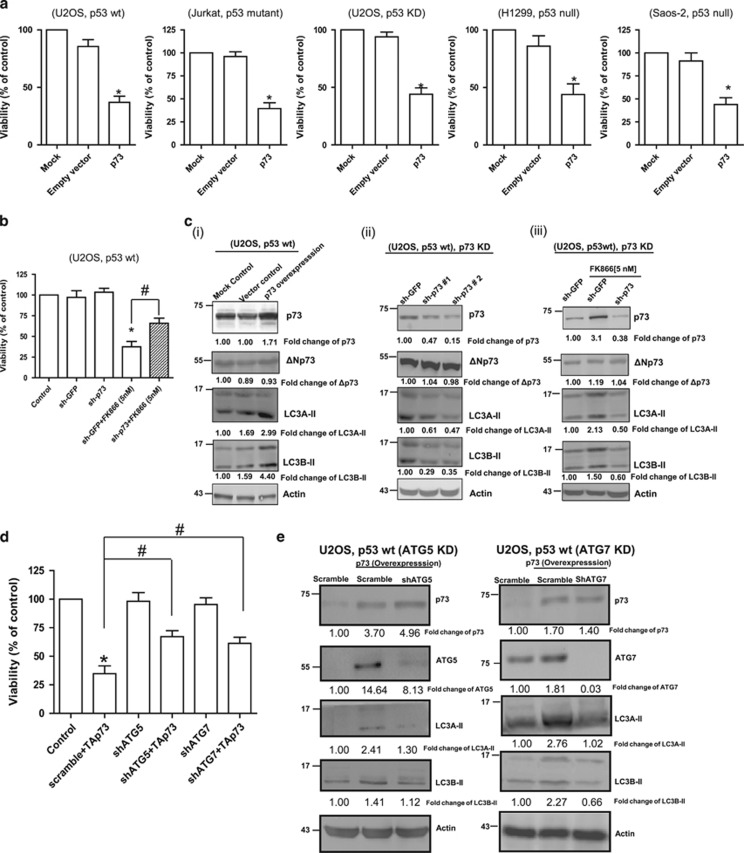
Figure 4.
Role of p73 in NAMPT inhibition- or NAMPT KD-mediated effect on autophagy and cell viability (cell count). (a) The various cell lines were transfected with p73 and cell viability was determined after 48 h using Trypan blue staining. (b) U2OS cells were transfected with either control (sh-GFP) or p73-specific shRNA (sh-p73), cultured in the presence or absence of FK866, and then analysed for cell viability after 48 h. (c) U2OS cells overexpressing p73 (i) or with p73 KD (ii) were analysed for basal levels of LC3A-II and LC3B-II; (iii) U2OS cells with control (sh-GFP) or p73 (sh-p73) KD were exposed to FK866 for 48 h and analysed for LC3A-II and LC3B-II. (d and e) Transient overexpression of p73 (for 48 h) was carried out in U2OS cells with or without ATG5 KD or ATG7 KD. Cell were then analysed for (d) viability, and (e) the levels of LC3A-II and LC3B-II. Statistical analysis for the cell viability study was performed with one-way analysis of variance (ANOVA) followed by paired ‘t'-test; *, #P-values ⩽0.05 obtained by comparing the respective data with the control. Results represent three independent experiments. sh, short hairpin; Wt, wild type

