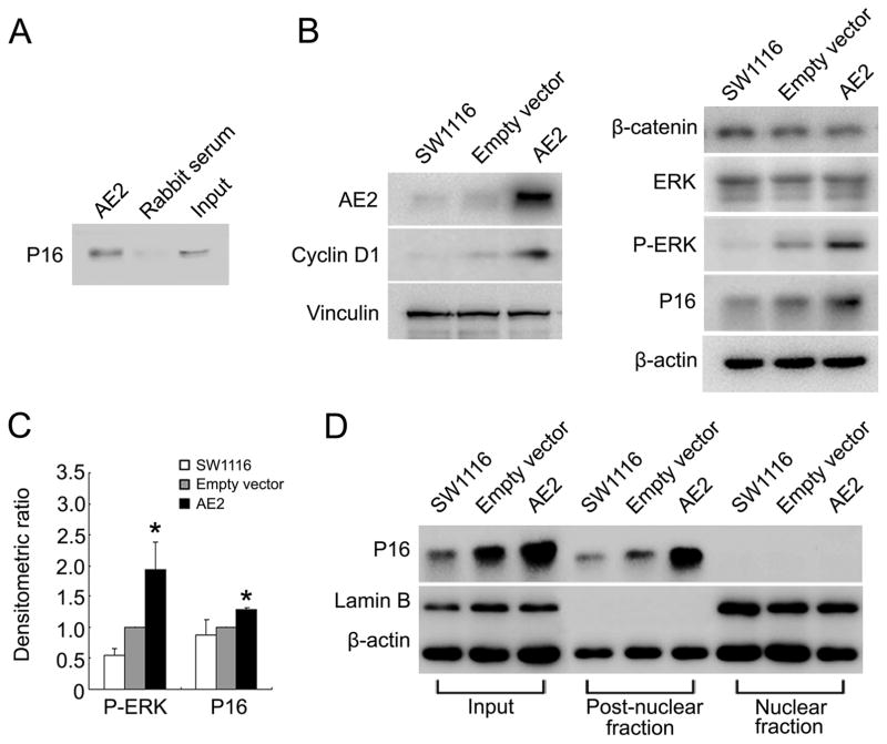
Figure 2. Interaction of AE2 and P16 in cytoplasm was associated with elevated P-ERK abundance in colon cancer cells.
(A) Interaction of AE2 with P16 in SW1116 cells. Cell lysates were subjected to immunoprecipitation with antibodies to AE2 or with rabbit serum. Immunoprecipitates were then immunoblotted with antibody to P16. (B) Immunoblot detection of cyclin D1, β-catenin, ERK, P-ERK and P16 proteins in SW1116 cells 24 hrs after transfection with pEGFP-AE2 or with pEGFP-C1 empty vector. (C) Densitometric scanning ratios of b-actin-normalized expression of P-ERK and P16 protein expression. Results were expressed as mean ± SD. *P<0.05 as compared with empty vector (n=3). (D) Nuclear and post-nuclear fractions of SW1116 cells were separated 24 hrs post-transfection with pEGFP-AE2 or pEGFP-C1 empty vector, then assessed for expression of P16 and Lamin B by immunoblot.

