
Figure 7.
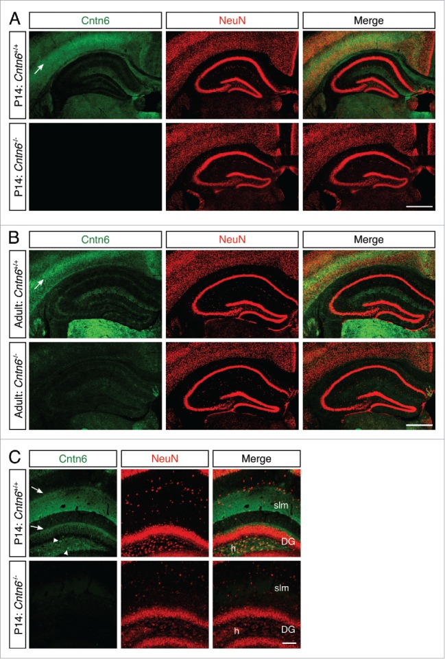
Cntn6 protein expression in the hippocampus. (A-B) Immunohistochemistry for Cntn6 (green) and NeuN (red) showed Cntn6 expression in the hippocampus of P14 and adult wild-type mice. No significant background staining was found in Cntn6−/− adult mice. Specific Cntn6-staining was also observed in the cortex of wild-type mice (arrows). (C) A higher magnification of Cntn6 (green) and NeuN (red) immunostaining of the hippocampus of P14 wild-type and Cntn6−/− mice. The arrowheads show Cntn6 expression at cell bodies in the hilus (h) of the dentate gyrus (DG) and the arrows indicate Cntn6 expression adjacent to the granule layer of the DG and at the stratum lacunosum-moleculare (slm) of wild-type P14 mice. All scale bars represent 100 μm.