Figure 2.
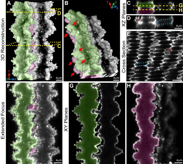
Confocal fluorescence microscopy images (2D single optical planes, extended focus flattened Z-stack, 3D reconstruction and cross section) of F-actin in neighboring mature fiber cells in 6-week-old Tmod1+/+ lenses. Single fiber cells are pseudocolored green or pink or uncolored. 3D reconstruction of Z-stacks through three neighboring fibers shows interlocking structures between adjacent cells (A) and coordinated paddle domains between cells of neighboring layers (B) (red arrows). Dashed boxes show the level where single XZ planes in (C, D) are derived. A single 2D XZ plane from the 3D reconstruction shows normal hexagonal fiber cell morphology (C, D) (hexagonal cell body outlined with blue dashed lines) with paddles and protrusions along the short side (D) (red asterisk). The F-actin staining pattern in dissociated fiber cells is similar to what is typically observed in cross sections through hexagonal mature lens fibers that have undergone denucleation (E) (hexagonal cell body outlined with blue dashed lines), demonstrating enriched F-actin along the short sides of these fibers and in regions of paddles and protrusions (red asterisk) with less intense F-actin signals along the broad sides. Extended focus image of the flattened Z-stack shows F-actin–rich interlocking paddles decorated by small protrusions along the short sides of the cells, creating a 3D zipper between adjacent cells (F). Single XY optical sections along the anterior–posterior axis through the fiber cells (G, H), where the dashed lines are drawn through the XZ plane in (C) show interlocking domains between fibers with some separation between the cells. Scale bars: 4 μm.

