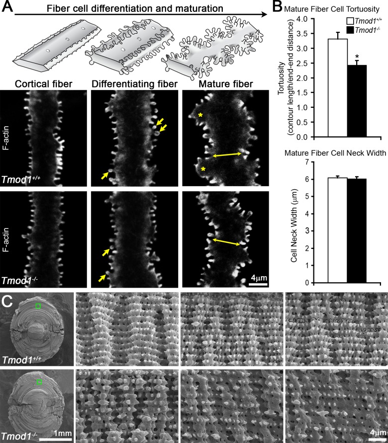
Figure 3.
Confocal fluorescence microscopy images (2D, single optical plane) of F-actin in fiber cells at various depths and SEM in 6-week-old (A) and 2-month-old (C) Tmod1+/+ and Tmod1−/− lenses. (A) Diagrams of normal cortical, differentiating, and mature fiber cells are shown along the top. Tmod1+/+ and Tmod1−/− cortical and differentiating fiber cells are straight with small F-actin protrusions along their vertices. Protrusions in differentiating fibers have more pronounced head and neck regions (arrows). Tmod1+/+ mature fibers have large paddle domains (asterisks) with F-actin–rich small protrusions, while Tmod1−/− fiber cells have F-actin–positive protrusions, but very few paddles. (B) Tmod1−/− mature fiber cells display a significant decrease in tortuosity (n = 9, *P < 0.01), but the cell neck width was unaffected (double-headed arrows in [A]) between Tmod1+/+ and Tmod1−/− lenses (n = 9). (C) Low-magnification SEM with boxed regions indicating the location of higher-magnification images. Comparable regions are identified by measuring outward from the nucleus at the center of the lens, since some peripheral cortical fibers are lost in the sample preparation procedure. Tmod1+/+ lenses have rows of mature fiber cells with coordinated paddle protrusions that are decorated by smaller protrusions of equal size and spacing. In contrast, Tmod1−/− mature lens fibers have disorganized paddles with irregular protrusions. Scale bars: 4 μm (A); 1 mm (C) (low magnification left); 4 μm (C) (high magnification).

