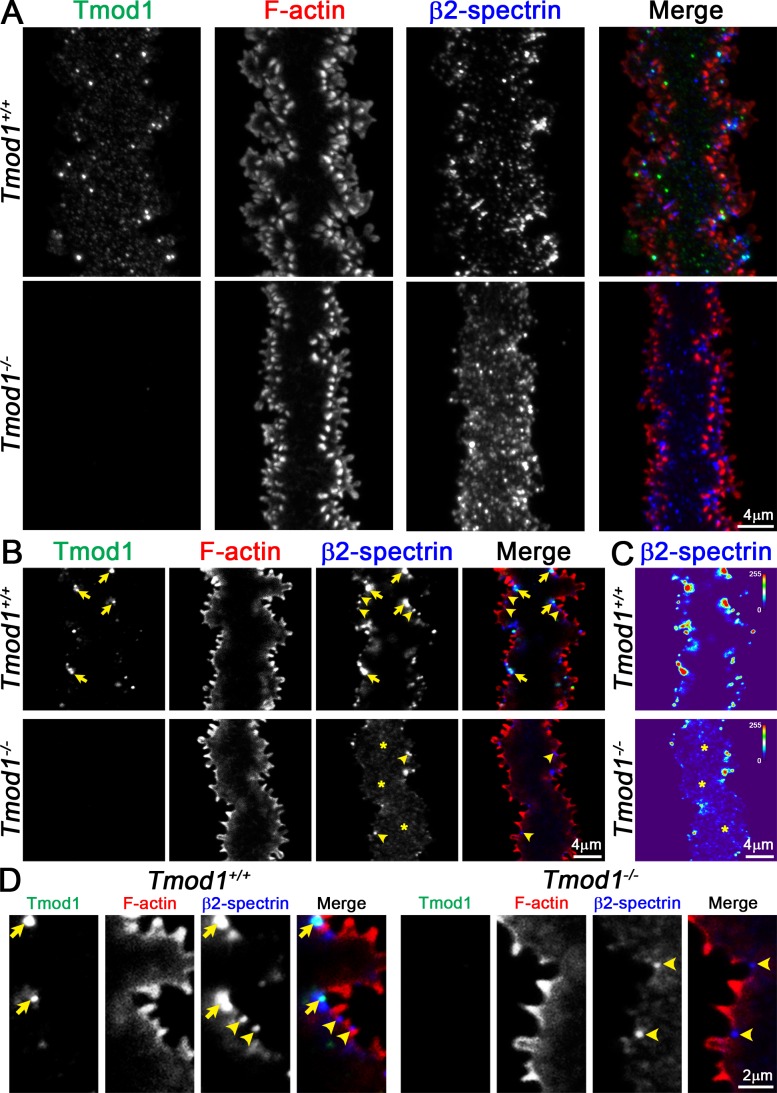
Figure 4.
Immunostaining of single mature fiber cells from 6-week-old Tmod1+/+ and Tmod1−/− lenses for Tmod1 (green), F-actin (red), and β2-spectrin (blue). (A) Extended focus of Z-stacks through mature fiber cells. The Tmod1+/+ fiber has F-actin–rich large paddle domains and small protrusions along cell vertices. Tmod1 and β2-spectrin are enriched in numerous puncta in the control fiber cell. While the Tmod1−/− fiber has very few paddles, F-actin–rich small protrusions are still present. (B, D) Single optical section (2D) from a Z-stack, showing a section through the cytoplasm of the fiber cells, with enlargements. Tmod1 and β2-spectrin are enriched in puncta near the cell membrane in valleys between large paddle domains in the Tmod1+/+ fiber (arrows), and β2-spectrin is also enriched at the base of small protrusions in Tmod1+/+ and Tmod1−/− fibers (arrowheads). The β2-spectrin staining signal appears diffuse and cytoplasmic (asterisks) with fewer membrane-associated puncta in the Tmod1−/− fiber. (C) Fluorescence intensity heat maps of β2-spectrin staining in Tmod1+/+ and Tmod1−/− lens fibers show that β2-spectrin staining is more cytoplasmic (asterisks) in the Tmod1−/− fiber. Scale bars: 4 μm (A–C); 2 μm (D).

