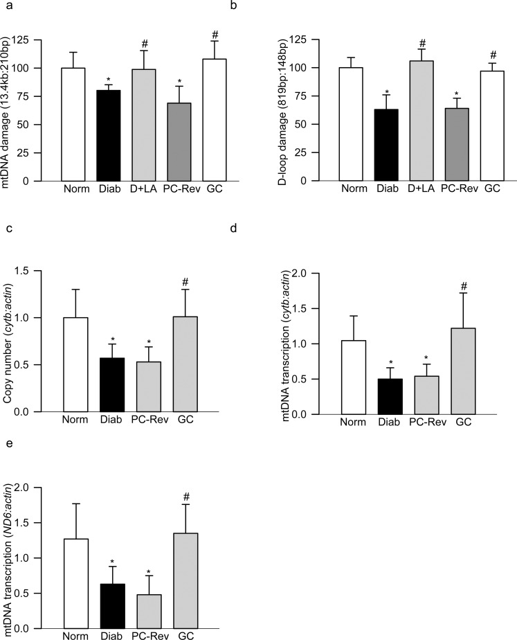
Figure 1.
Mitochondrial DNA damage in peripheral blood of diabetic rats. DNA damage was determined by amplifying the (a) 13.4-kb and 210-bp amplicons of the mtDNA, and (b) 819-bp and 148-bp amplicons in the D-loop region of the mtDNA. The relative amplification was quantified by normalizing the intensity of the long PCR product to the short PCR product. Decrease in the amplification ratio indicated an increase in the DNA damage. (c) Copy numbers of mtDNA were determined in the genomic DNA by qPCR using cytb as mtDNA marker and β-actin as a nuclear DNA marker. Transcripts of mtDNA-encoded (d) cytb and (e) ND6 were quantified by SYBR green–based qPCR using β-actin as a housekeeping gene. Each measurement was made in duplicate in five to seven rats in each group, and the values are represented as mean ± SD. Norm, normal; Diab, diabetes; PC-Rev, poor control for 6 months followed by good control for 6 months; GC, good glycemic control. *P versus normal and #P < 0.05 versus diabetes.

