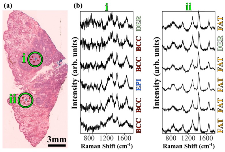
Fig. 7.
Example multiplex Raman measurements and diagnosis for a typical skin tissue sample from surgery. (a) Adjacent H&E tissue section, with the estimated measurement locations shown with green circles and manually selected sampling pattern (scaled up in size for clarity, not representative of exact sampling locations). (b) 6-beam multiplex Raman spectra (with baseline subtracted) from the labelled regions in the bright-field image, with corresponding diagnoses. Total laser power at sample: 1W; integration time: 2 seconds (batches of 6 sampling points). Spectra are shifted vertically for clarity.

