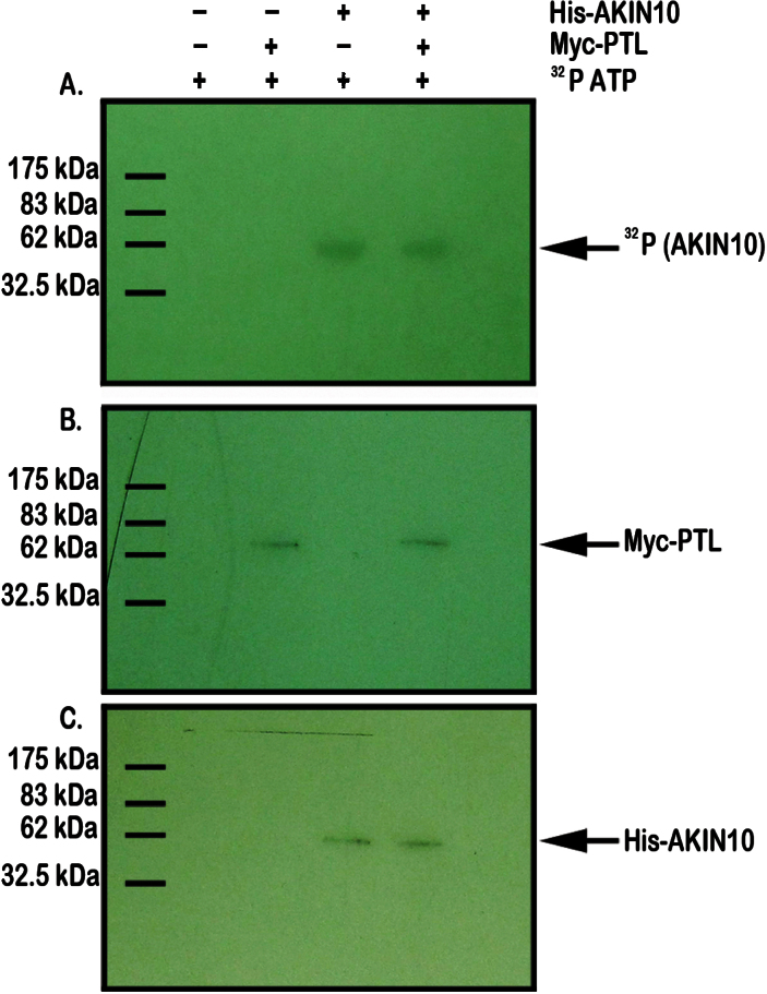
Fig. 6.
Test of phosphorylation of PTL by AKIN10. Mixed extracts of epitope-tagged AKIN10 and PTL proteins were incubated with [γ-32P]ATP and separated by gel electrophoresis. (A) Autoradiograph showing 32P-labelling of His–AKIN10-sized bands in lanes 3 and 4, but no labelling of Myc–PTL-sized bands in lanes 2 and 4, indicating that AKIN10 alone has been phosphorylated. (B) Western blot of the same extracts probed with anti-Myc antibody showing the presence of tagged PTL protein in lanes 2 and 4. (C) Western blot of the same extracts probed with anti-His antibody showing the presence of tagged AKIN10 protein in lanes 3 and 4.

