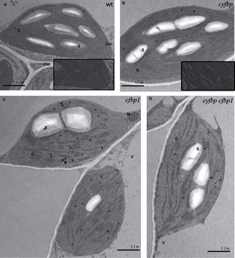
Fig. 5.
Transmission electron microscopy analysis of leaf sections from wild- type (wt) (A), cyfbp1 (B), cfbp1 (C), and cyfbp cfbp1 (D) plants. Leaves were collected at 4h in a 16h light/8h dark photoperiod, fixed, embedded, and sectioned as described in the Materials and methods. G, grana; S, starch; V, vacuole; P, plastoglobule; Chl, chloroplast; CW, cell wall; M, mitochondrion.

