Fig. 6.
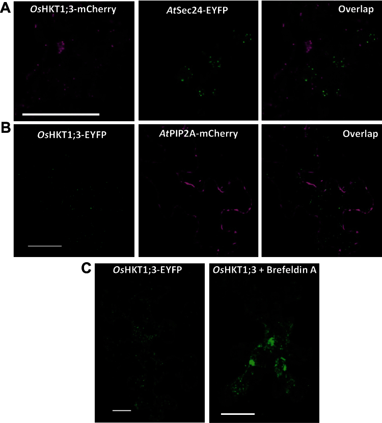
OsHKT1;3 does not localize to the ERES or the plasma membrane and forms aggregates upon exposure to brefeldin A. (A) Expression of OsHKT1;3–mCherry (left) and the ERES/COPII marker AtSec24–EYFP (centre), and overlapping of the two images (right). (B) Co-localization analysis of OsHKT1;3–EYFP (left) with the plasma membrane marker AtPIP2A–mCherry (centre), and overlapping of the two images (right). The intracellular localization of OsHKT1;3–EYFP, seen as fluorescent puncta distributed throughout the cell (C, left), was modified after incubation of the epidermis with brefeldin A at 25 μM for 15min, resulting in the formation of aggregated bodies (C, right). Scale bar=25 μm.

