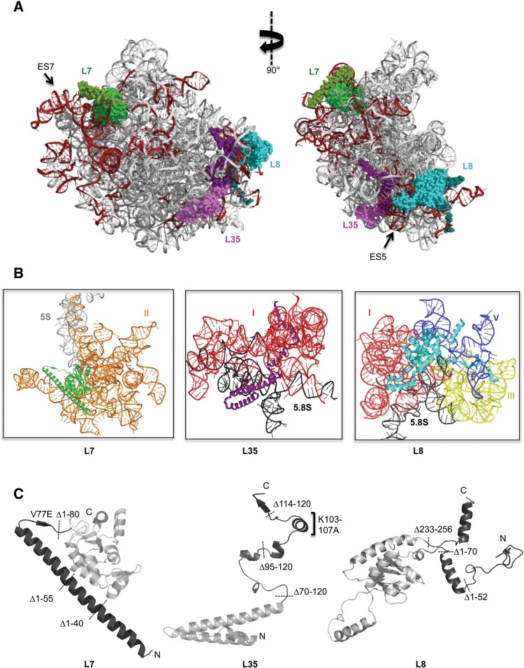
FIGURE 1.
(A) Positions of r-proteins L7 (green), L35 (purple), and L8 (cyan) are shown in the mature yeast 60S ribosomal subunit. Eukaryote-specific extensions of r-proteins are indicated in darker shades of the corresponding colors. Expansion segments of 25S rRNA are colored dark red. The structure on the left corresponds to the solvent-exposed side of the 60S ribosomal subunit of S. cerevisiae (PDB 3U5H and 3U5I). (B) Cartoon representations of r-proteins L7, L35, and L8, showing the 25S rRNA domains that are contacted by globular regions and extensions. Domains I (red), II (orange), III (yellow), and V (blue) are indicated. (C) Globular domains of the proteins are indicated in light gray and extensions are shown in dark gray. Positions of the truncation mutations, and important point mutations investigated in this study are shown.

