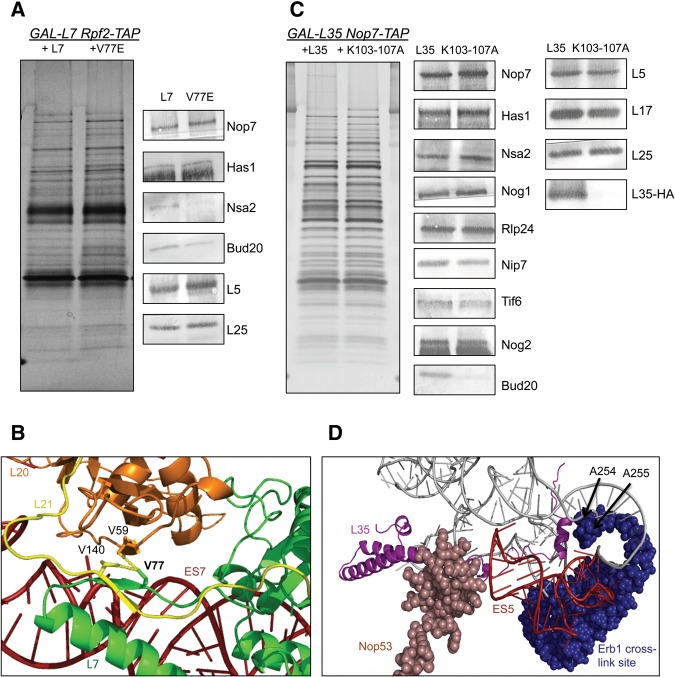
FIGURE 4.
(A) TAP-tagged assembly factor Rpf2 was used as the bait to assay the protein composition of pre-ribosomes in the L7V77E mutant strain. Protein levels were initially tested by SDS-PAGE, followed by silver staining (left panel). Antibodies against the indicated proteins were used to test their abundance by Western blotting. (B) Zoomed-in representation of the hydrophobic interactions mediated by the V77 residue of L7 (green), the V140 residue of L21 (yellow), and the V59 residue of L20 (orange). PyMol generated from the cryo-EM structure of Nog2 containing pre-ribosomes (PDB accession number 3JCT). (C) TAP-tagged assembly factor Nop7 was used as the bait to assay the protein composition of pre-ribosomes in the L35K103-107A mutant strain. Protein levels were initially tested by SDS-PAGE, followed by silver staining (left panel). Antibodies against the indicated proteins were used to test their abundance by Western blotting. (D) Zoomed-in representation of the C-terminal extension of L35 (purple). Assembly factor Nop53 was colored light pink while the cross-linking site of assembly factor Erb1 is shown in dark blue. ES5 is indicated in red. PyMol generated from the cryo-EM structure of Nog2 containing pre-ribosomes (PDB accession number 3JCT).

