FIGURE 6.
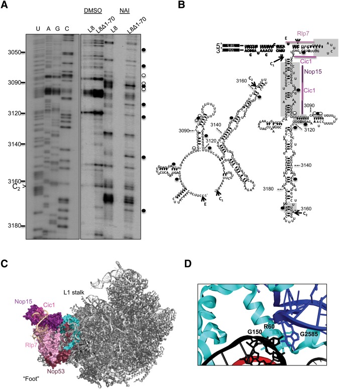
(A) In vivo structure probing of rRNA, using NAI followed by primer extensions, was conducted for L8 wild-type and L8Δ1-70 mutant yeast. Nucleotides with increased modification in the mutant are marked with solid black circles, while hollow circles indicate the nucleotides with decreased modification. (B) Changes in the nucleotide reactivity are shown in the ring conformation and the hairpin conformation of ITS2. The gray boxes correspond to structures necessary for progression of 60S subunit assembly (Côté et al. 2002). The assembly factors that interact with the first 59 nt of ITS2 in the cryo-EM structure of Nog2-associated particles were indicated on the hairpin conformation (PDB accession number 3JCT). (C) The position of L8 (cyan) in Nog2-associated pre-60S ribosomal subunits is shown from the subunit interface. The proteins that bind the “foot” structure are colored different shades of pink. ITS2 is colored beige. (D) The R60 residue in the N-terminal extension of L8 interacts with both 5.8S rRNA in the proximal stem (nucleotide G150) and domain V (nucleotide G2585) of 25S rRNA.

