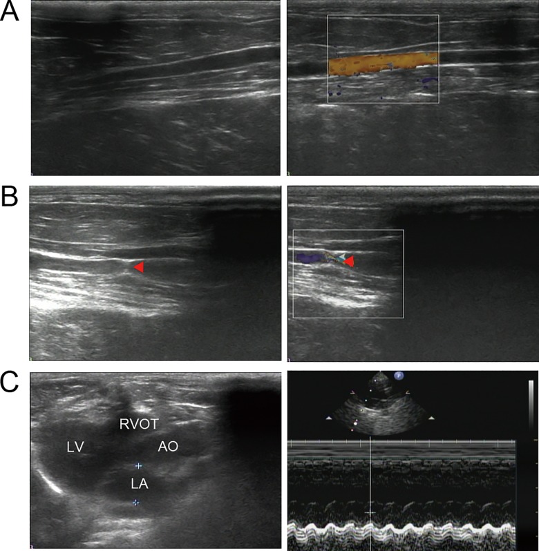
Fig 1. Color Doppler Ultrasound evaluation of abdominal aortic stenosis and cardiac structure of studied animals.
The gray-scale sonographic appearance (the left) and the color doppler flow imaging (the right) of the abdominal aorta for sham-operated (A) and AAC (B) rabbits. Red triangle indicated the position of the abdominal aortic stenosis. (C) Representative echocardiographic image with two-dimensional parasternal long-axis view and M-mode echocardiogram for a studied rabbit. AAC, abdominal aortic constriction; LV, left ventricle; LA, left atrium; AO, aorta; RVOT, right ventricular outflow tract.

