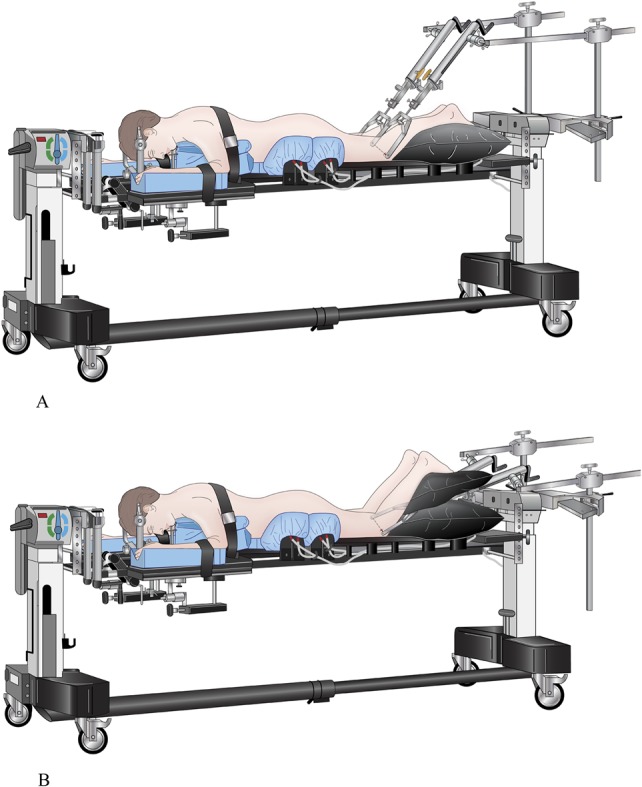
FIGURE 1.

Drawing of patient positioning on the OSI spine table. The head is secured with Mayfield head tongs and the patient is in bifemoral skeletal traction. A, The traction bows are placed posterior to the thighs in order to produce pelvis extension to reduce a sacral fracture kyphotic deformity. B, The traction bows are placed anterior to the thighs in order to produce longitudinal traction to reduce sacral fracture shortening as occurs in traumatic spondyloptosis as seen in Figure 4.
