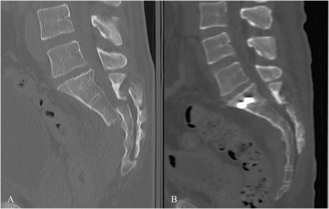
FIGURE 4.

These are images from the patient shown in Supplemental Digital Content 7 with traumatic spondyloptosis. A, Shows the preoperative sagittal CT scan, with complete dorsal displacement and shortening. B, Immediate postoperative CT scan showing anatomic reduction of the fracture. The iliosacral screws are seen in the S1 body. The percutaneous lumbopelvic fixation is in place but is not seen because the implants are all out of the plane of the image.
