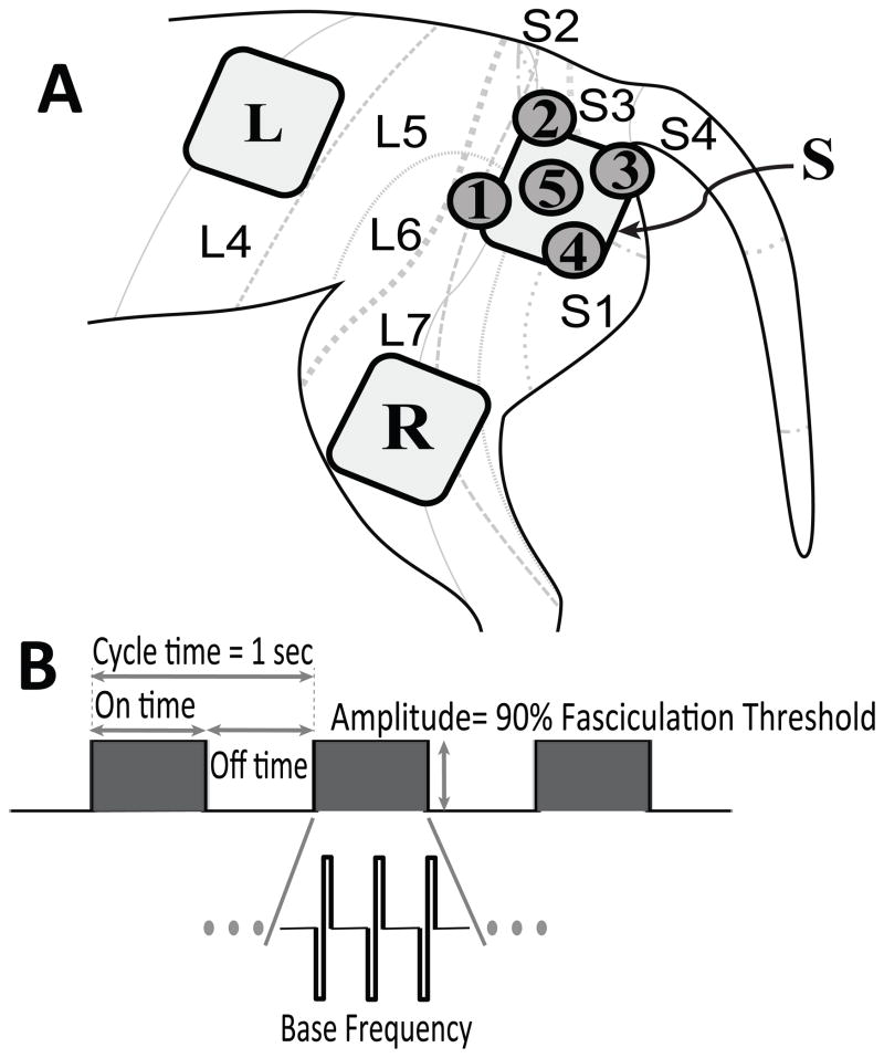
Figure 1. Surface electrode placement and patterned stimulus waveform.
A: Large 4 cm square electrode patches were used to stimulate sacral dermatomes [S] (L7–S3) and lumbar dermatomes [L] (L4–L5). Smaller 2 cm round patches were used to improve spatial resolution within the sacral dermatomes [1–5]. Return patch [R] for anodal stim was used for all electrodes. Feline dermatome map adapted from Kuhn (22), showing the approximate boundaries and overlap of L4–S4 dermatomes. Electrode [S] overlaps the L7–S3 dermatomes; [1]: L5–L7; [2]: L7/S2; [3]: L7–S1; [4]: S3; [5]: L7–S2 dermatomes.
B: The stimulus waveform was defined in terms of ON/OFF time, base frequency, and amplitude. On/ off times always summed to a total cycle time of 1 sec. Base frequency was fixed at 20 Hz for duty cycles less than 100%; other base frequencies were tested for continuous stimulation.

