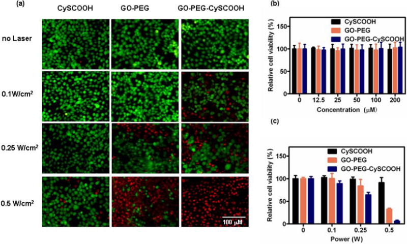
Figure 2.

In vitro cell experiments. a) Fluorescence image of Calcein AM/PI co-stained 4T1 cells incubated with 89.7 μg/ml of free CySCOOH, 160 μg/ml of GO-PEG and 250 μg/ml of GO-PEG-CySCOOH (equal to CySCOOH concentration of 100 μM) for 24 h after laser irradiation at different power densities (0.1, 0.25 and 0.5 W/cm2) for 10 min. The cells incubated with same concentration of free CySCOOH, GO-PEG and GO-PEG-CySCOOH without laser irradiation as control group (Scale bars: 100 μm). b) Relative viability of 4T1 cells incubated with various concentrations of free CySCOOH, GO-PEG or GO-PEG-CySCOOH for 24 h .c) Relative viability of 4T1 cells incubated with various concentrations of free CySCOOH, GO-PEG or GO-PEG-CySCOOH after 808- nm laser irradiation at different power densities (0.1, 0.25 and 0.5 W/cm2) for 10 min.
