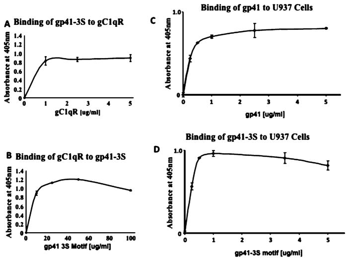
Fig. 5.
Interaction between HIV-1 gp41-3S and gp41 and recombinant gC1qR and cell surface expressed gC1qR.
(A) Duplicate wells of a microtiter plate were coated with 100 μl of 2 μg/ml gC1qR and incubated (60 min, 37C). After blocking, the bound gC1qR was reacted (1 h, 37 °C) with concentrations of synthetic gp41 3S peptide ranging from 1 to 5 μg/ml (A) and the bound peptide was detected using an affinity purified rabbit anti-gp41 peptide IgG. In (B) the reaction sequence was reversed in that the wells were first coated with 100 μl of 100 μg/ml gp41-3S peptide, then was reacted with increasing concentrations of biotinylated gC1qR (0–100 μg/ml) and the bound gC1qR was detected using alkaline phosphatase conjugated streptavidin. The experiments in (C) and (D) are similar except that microtiter plate-fixed cells were used and reacted with either various concentrations of purified gp41 (C) or the gp41-3S peptide (D) and the bound protein was detected using mAb to gp41 or immunoaffinity purified anti-gp41-3S peptide antibody. Each data point is a mean ± SD run in duplicates (n = 3).

