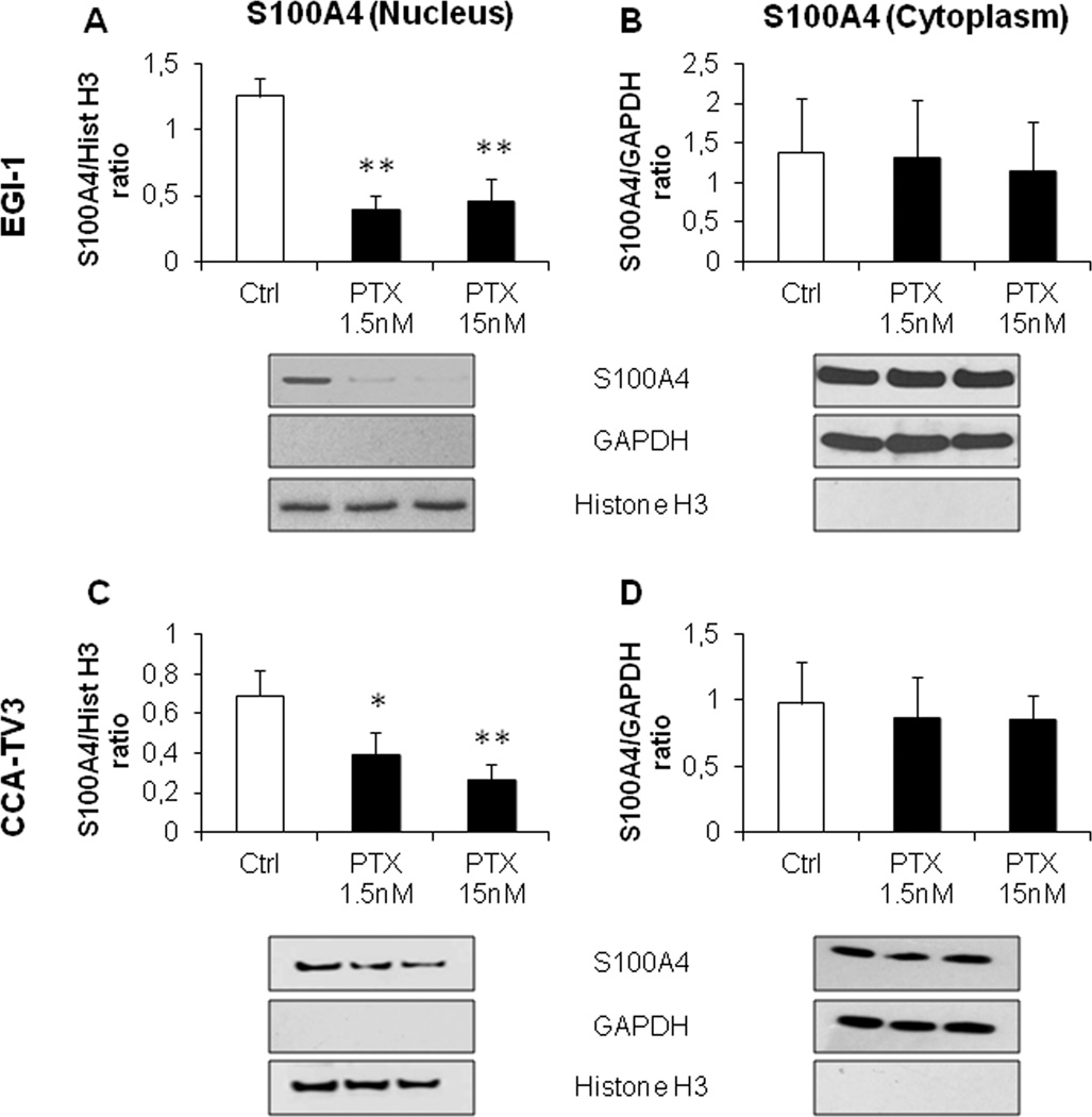
Figure 1. Low dose PTX reduced nuclear but not cytoplasmic S100A4 expression in established (EGI-1) and primary (CCA-TV3) CCA cell lines.
A–D. Treatment with PTX at low doses (1.5, 15nM) induced a significant reduction in the nuclear (A,C) but not in the cytoplasmic S100A4 content (B,D), with respect to controls, in both cell lines. Below each column plot, representative blots of S100A4 together with histone H3 and GAPDH (markers of nuclear and cytoplasmic fractions, respectively), are shown (n=5 for EGI-1; n=3 for CCA-TV3). *p<0.05 vs Ctrl, **p<0.01 vs Ctrl.

