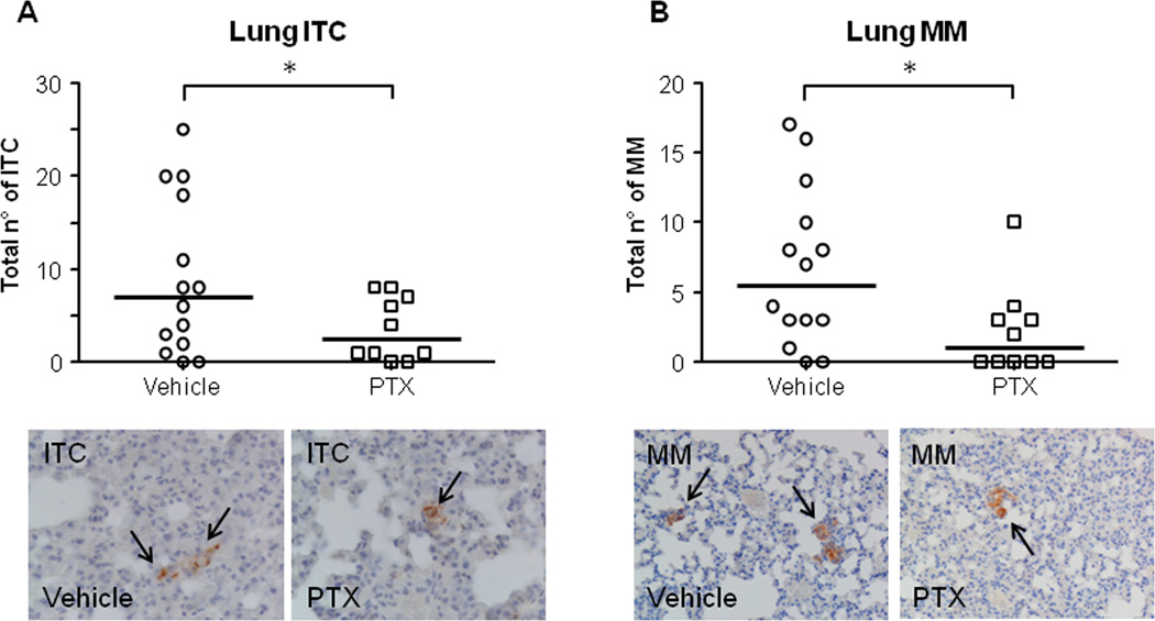
Figure 7. LDM PTX inhibited lung metastatic colonization of EGI-1 cells, in vivo.
A–B. A significant reduction in the number of both ITC (A) and MM (B) was found in treated mice (n=10) with respect to controls (n=14). Representative micrographs of ITC and MM (black arrows) derived from EGI-1 cell dissemination to the lung parenchyma after xenograft in SCID mice undergoing LDM PTX and in controls, identified by the specific immunoreactivity for human mitochondria, are shown below their respective dot plot. *p<0.05 vs Ctrl; Immunoperoxidase; original magnification, M=400× (ITC), M=200× (MM).

