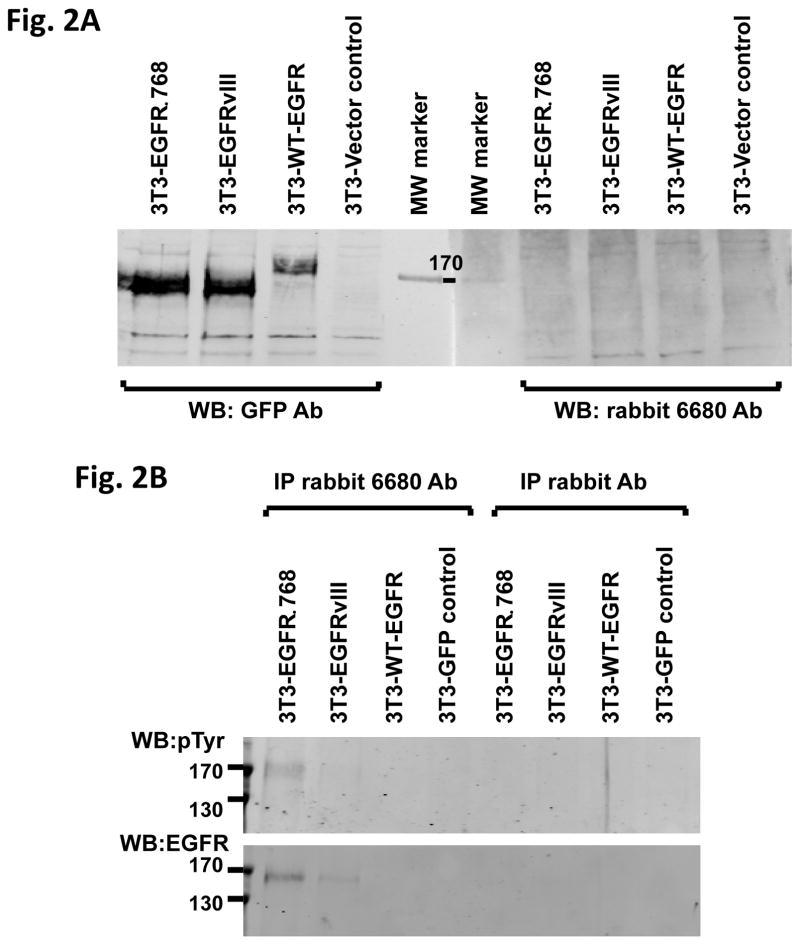
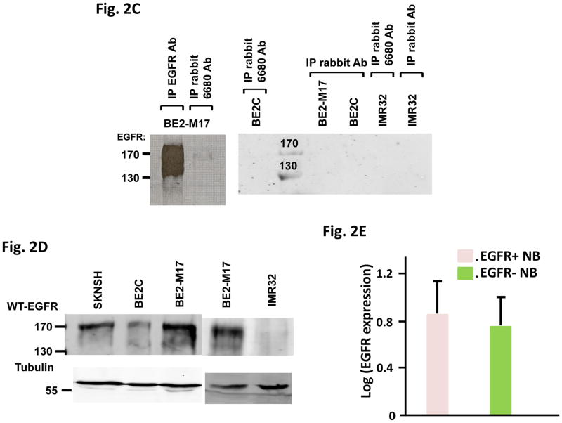
Fig. 2. EGFRΔ768 protein expression in NB.
(A) Western blot analysis of 3T3 cell lysates expressing GFP (vector control), WT-EGFR-GFP, EGFRvIII-GFP and EGFRΔ768-GFP using the purified EGFRΔ768 polyclonal rabbit antibody (6680 Ab) and GFP antibody. MW: molecular weight. (B) Immunoprecipitation followed by western blotting (IPW) analyses of 3T3 cell lysates expressing GFP, WT-EGFR-GFP, EGFRvIII-GFP and EGFRΔ768-GFP using the EGFRΔ768 specific polyclonal rabbit antibody (6680 Ab). The negative controls were IP with rabbit IgG antibody. (C) EGFRΔ768 protein expression in the BE2M17 cell line. BE2M17, BE2C and IMR32 cell lysates were analyzed by IPW using the EGFRΔ768 specific polyclonal rabbit antibody (6680 Ab). The negative controls were IP with rabbit IgG antibody (lane #3, 4 and 6 of the right blot). The positive control was IP with the EGFR antibody, cetuximab (lane #1 of the left blot). (D) WT-EGFR expression in a panel of NB cell lines. (E) Comparison of WT-EGFR expression between ΔEGFR+ (n = 17) and ΔEGFR- (n = 42) CBRC primary NB tumors. ΔEGFR = EGFRvIII + EGFRΔ768. EGFR expression was quantified and compared as previously described (11). 3 of the 20 ΔEGFR+ CBRC NB tumors did not have enough materials for protein analysis. Error bars represent ±2 standard errors.


