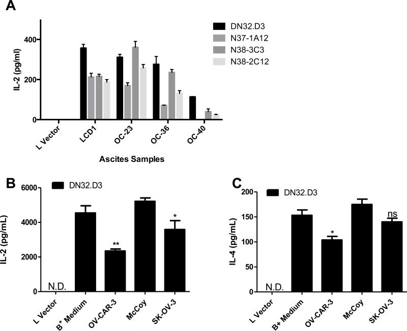
Figure 1. Tumor Ascites and Conditioned medium from ovarian cancer cell lines inhibits NKT cell activation.
(A) LCD1d cells were treated with control medium or with ovarian cancer ascites fluid from patients for 4 h, then washed extensively and cocultured with a panel of NKT cell hybridomas (DN32.D3, N37-1A12, N38-3C3 and N38-2C12). After 20-24 h, IL-2 was measured as an indication of NKT cell activation using standard cytokine ELISA. (B, C) LCD1d cells were treated with supernatants from confluent ovarian cancer cell lines OVCAR-3 and SK-OV-3 for 4 hours at 37°C and washed extensively following treatment. Control LCD1d cells were concurrently treated with RPMI (B+ Medium) and McCoy media. Lvector cells serve as a negative control. N.D.=not detectable. Following treatment, LCD1d cells were co-cultured with NKT cell hybridomas, DN32.D3, and incubated for 24 hours at 37° C. Standard ELISA was performed to measure cytokine production (B) IL-2 (C) IL-4. Data are shown as mean ±S.E.M. of one experiment set up in triplicate. The experiments were performed at least twice with each ascites sample and three times with conditioned medium. T-tests were performed to compare medium vs. conditioned medium groups, yielding p-value of p=0.0061 for IL-2 and p=0.0187 for IL-4.

