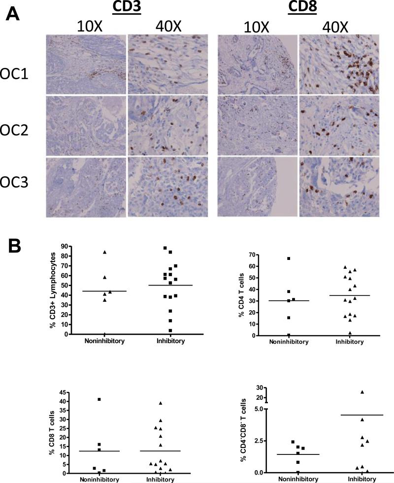
Figure 2. Lymphocytes are present within the tumor microenvironment.
(A) Representative images of patient tissue sections from confirmed cases of ovarian cancer were stained for CD3 and CD8. (B) Flow cytometry was performed to assess T cell subpopulations within the ascites. CD1d-expressing cells were treated with cell-free ascites for 4 hours, then washed extensively and cocultured with a panel of NKT cell hybridomas. Samples that caused <10% inhibition of IL-2 production by NKT cells were deemed non-inhibitory, the others are referred to as inhibitory.

