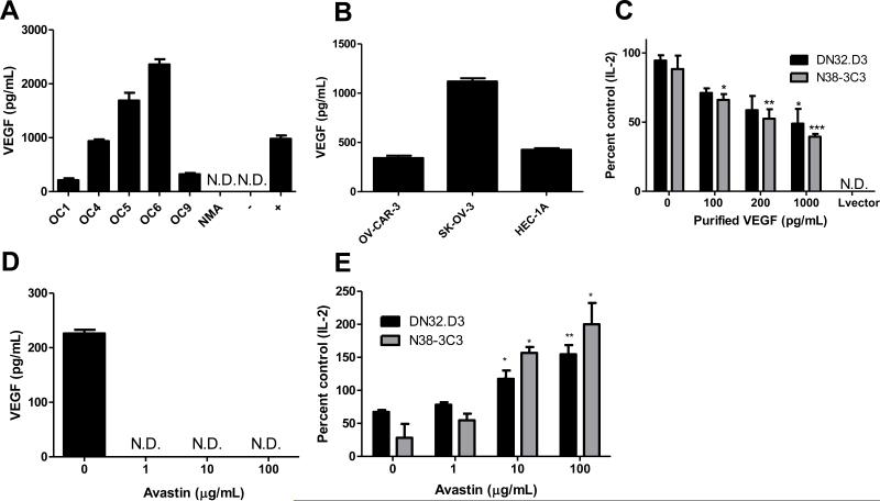
Figure 3. VEGF inhibits NKT cell function.
(A) VEGF levels in ovarian cancer patient ascites and (B) OV-CAR-3, SK-OV-3 and endometrial cancer cell lines HEC-1 were measured by ELISA. In (A), non-malignant ascites (NMA) represents VEGF levels in non-cancer-associated ascites. The negative control is the assay diluent used in the ELISA assay and the positive control was recombinant VEGF (1000 pg/mL). ND=not detectable. (C) LCD1d cells were treated with the indicated concentrations of recombinant VEGF for 4 hours, then washed extensively and cocultured with NKT cell hybridomas, DN32.D3 and N38-3C3. (D) OV-CAR-3 cells were treated with increasing concentrations of Bevacizumab/Avastin and VEGF levels in the cell culture supernatant were assessed after 24 hours by ELISA. (E) OV-CAR-3 cells were treated with increasing concentrations of Bevacizumab/Avastin for 3 days, following a one-day recovery period in fresh culture medium. Pretreatment of CD1d-expressing cells with conditioned medium were performed as described above. ANOVA with Bonferroni post-test confirmed significance. *p<0.05; **p<0.01; and ***p<0.001 for treatment groups compared to control.

