
Figure 3.
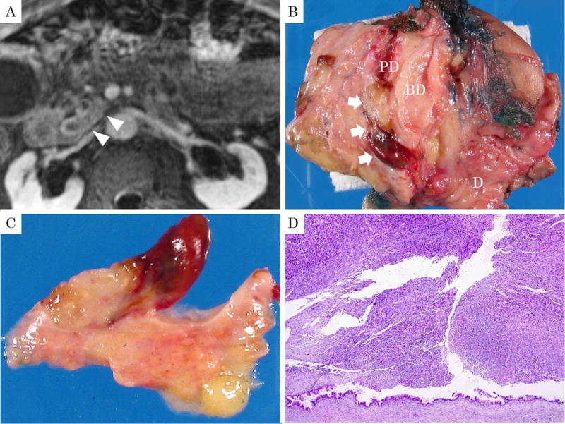
86-year-old, male with undifferentiated carcinoma with osteoclastic giant cells. A: Magnetic resonance image revealed a tumor within the dilated main pancreatic duct (arrowheads) along with thickening of pancreatic duct wall on delay phase of T1-weighted image with fat suppression. B, C: Macroscopically, the polypoid tumor projected into the dilated pancreatic duct (arrow). The polyp was soft and yellow with hemorrhagic foci and a thin stalk. (BD: common bile duct, PD: main pancreatic duct, D: duodenum) D: Macroscopic appearance of the same polypoid lesion showing sheet-like growth of sarcomatoid tumor with characteristic OGC pattern. Preserved duct epithelium is seen at the base and shows low-grade PanIN. (H&E stain, ×40)