
Figure 7.
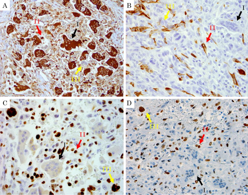
Immunohistochemical findings of undifferentiated carcinoma with osteoclastic giant cells. Black arrow I: Osteoclastic cells. Red Arrow II: Histiocyte-like sarcomatoid carcinoma cells (HSCs). Yellow arrow III: Pleomorphic/undifferentiated malignant cells (PCs). A: Immunolabelling for CD68 (lysosome, macrophage marker) labels osteoclastic cells strongly, and also marks HSCs albeit more weakly. Note that pleomorphic/undifferentiated malignant cells are being engulfed by the osteoclastic cells are not stained. (CD68, ×400) B: In contrast, AE1/AE3 is negative in osteoclastic cells and HSCs, but show focally positivity in HSCs and PCs (AE1/AE3, ×400) C, D: HSCs and PCs had a high expression of p53 and a high proliferation rate with Ki-67 stain, while osteoclastic cells were negative for both. (C: Ki-67, ×400, D: p53, ×400)