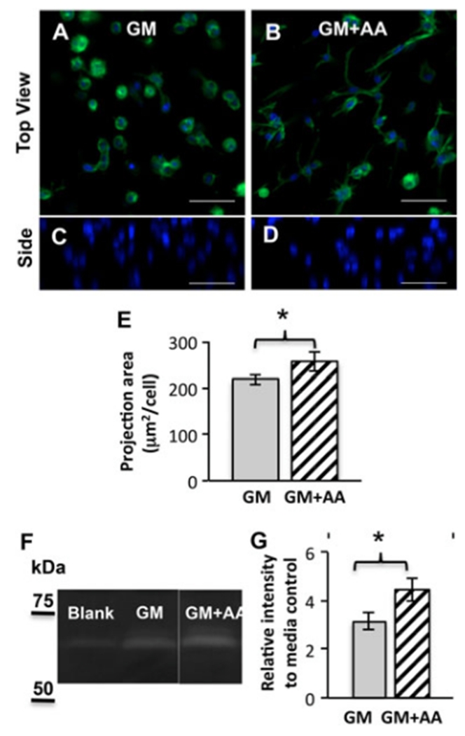
Figure 2.
AA promotes cell spreading and MMP-2 secretion within MMP-labile PEG–PQ hydrogels. (A–D) Representative images of cell spreading within hydrogels after encapsulation for 1 day in (A, C) GM or (B, D) GM + AA; blue (DAPI), nuclei; green (phalloidin), actin; top (A, B) and side (C, D) views; scale bars=50μm. (E) Average z-projection area of VIC spreading within hydrogels at day 1; *p < 0.05; n = 3. (F–G) Zymography of fresh media control and conditioned media collected from encapsulated VIC culture (days 1–4): (F) Representative image of zymography with clear bands corresponding to MMP-2; (G) Quantitative analysis of band intensities relative to the media control; *p<0.01; n=4–5

