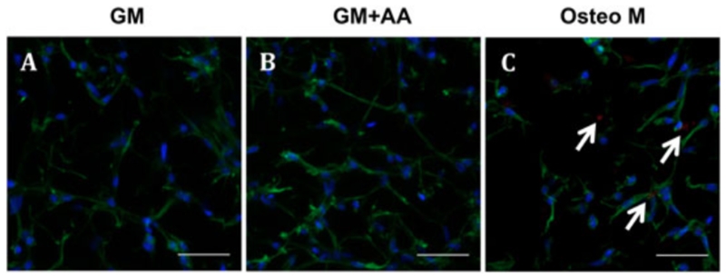
Figure 6.
Xylenol orange (calcification) was undetected in cell-laden hydrogels cultured in GM or GM + AA for 28 days, but was faintly detectable in those cultured in Osteo M: red (xylenol orange), calcium; blue (DAPI), nuclei; green (phalloidin), actin: representative fluorescent images of cell-laden hydrogels cultured in (A) GM, (B) GM + AA and (C) Osteo M; scale bars = 50 μm; white arrows, calcification

